

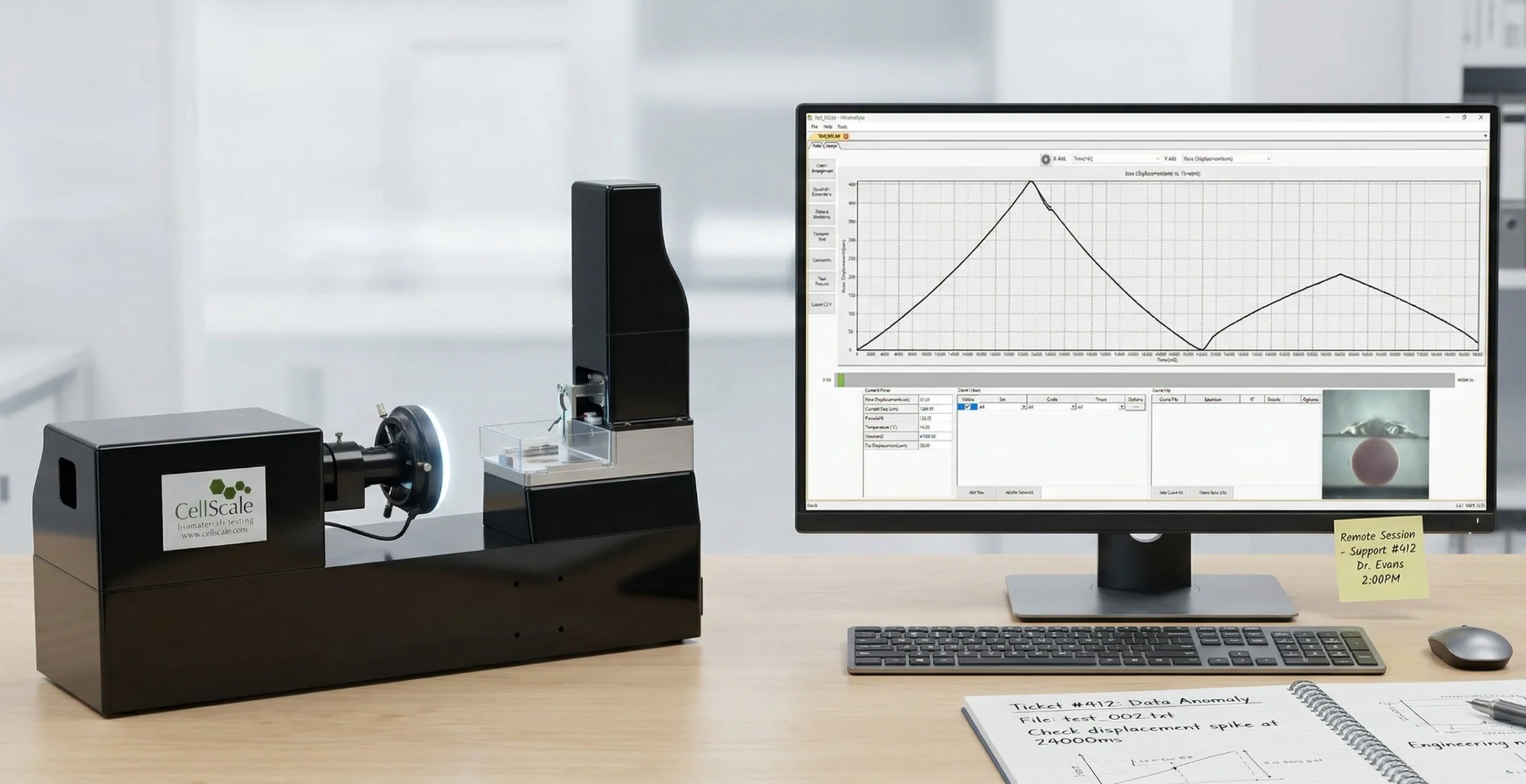


MicroTester Videos
5 Videos
4:01

3:26
2:36

3:29

7:28
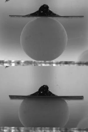
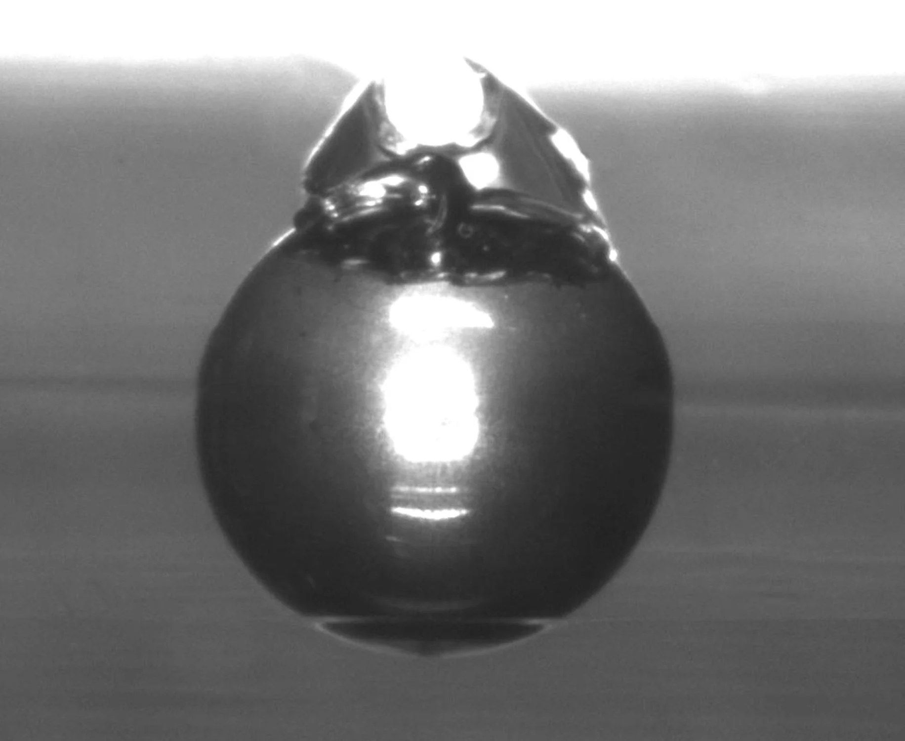
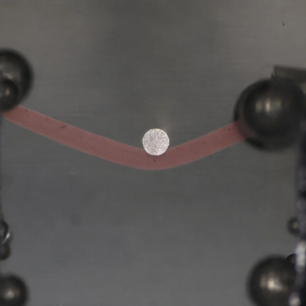
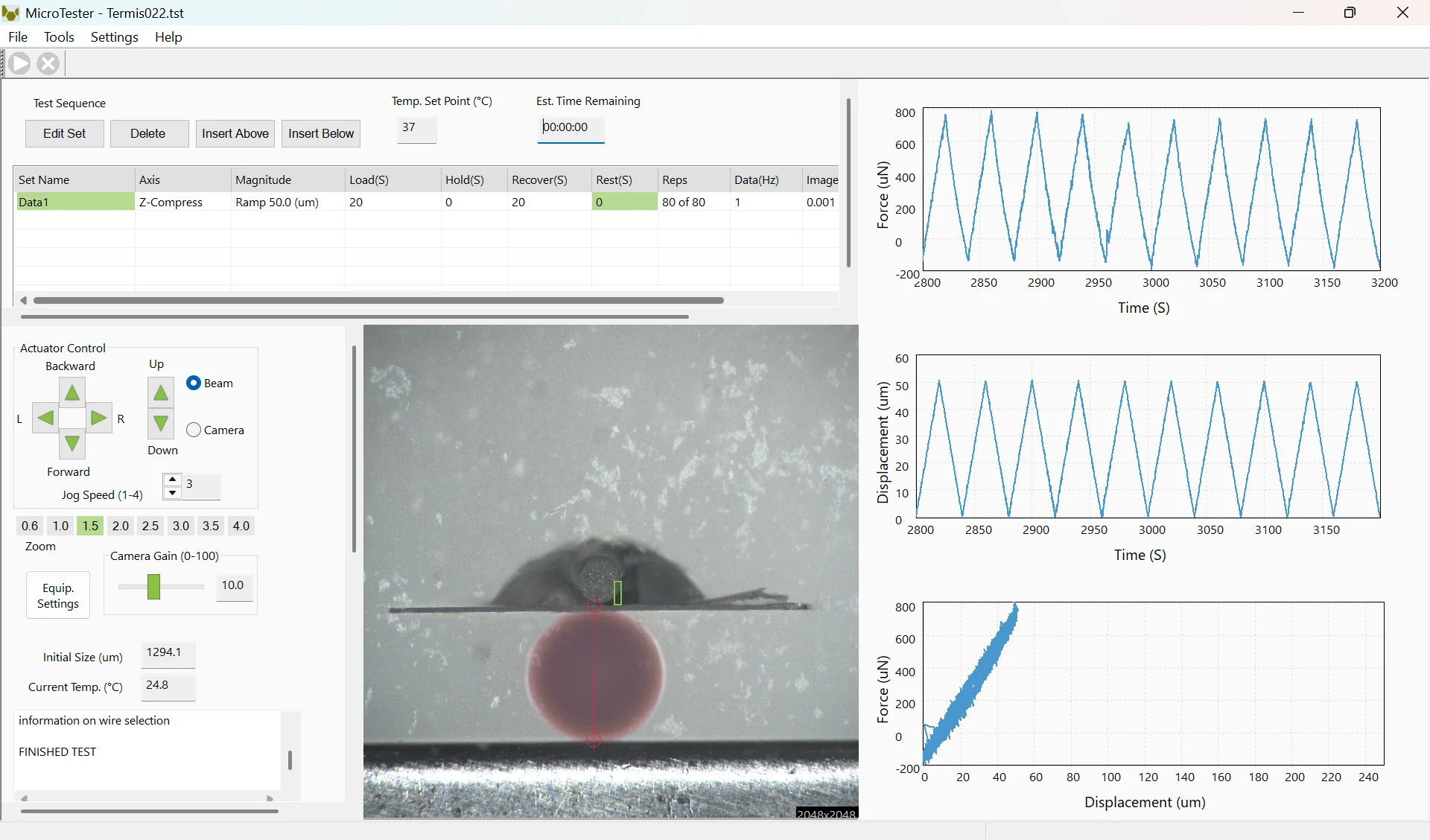
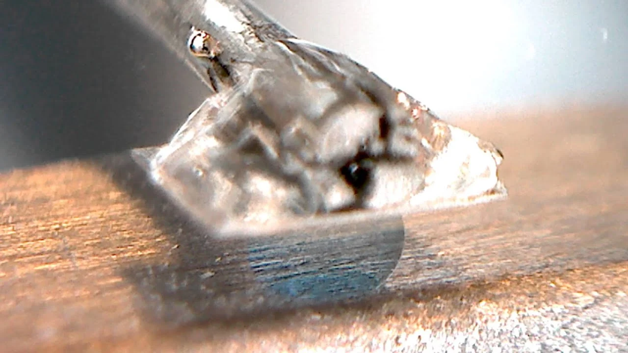
Hydrogel Microsphere Parallel-Plate Compression Test Demonstration
Step-by-step demonstration of parallel-plate compression testing on hydrogel microspheres using the MicroTester.
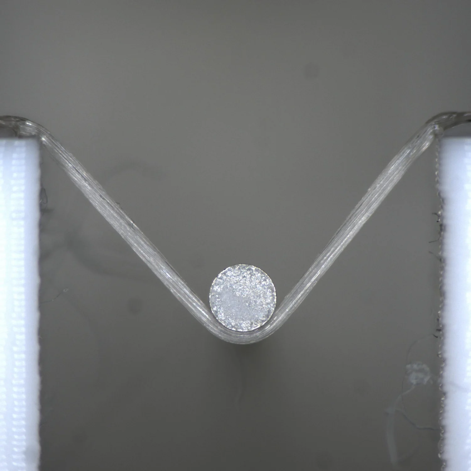
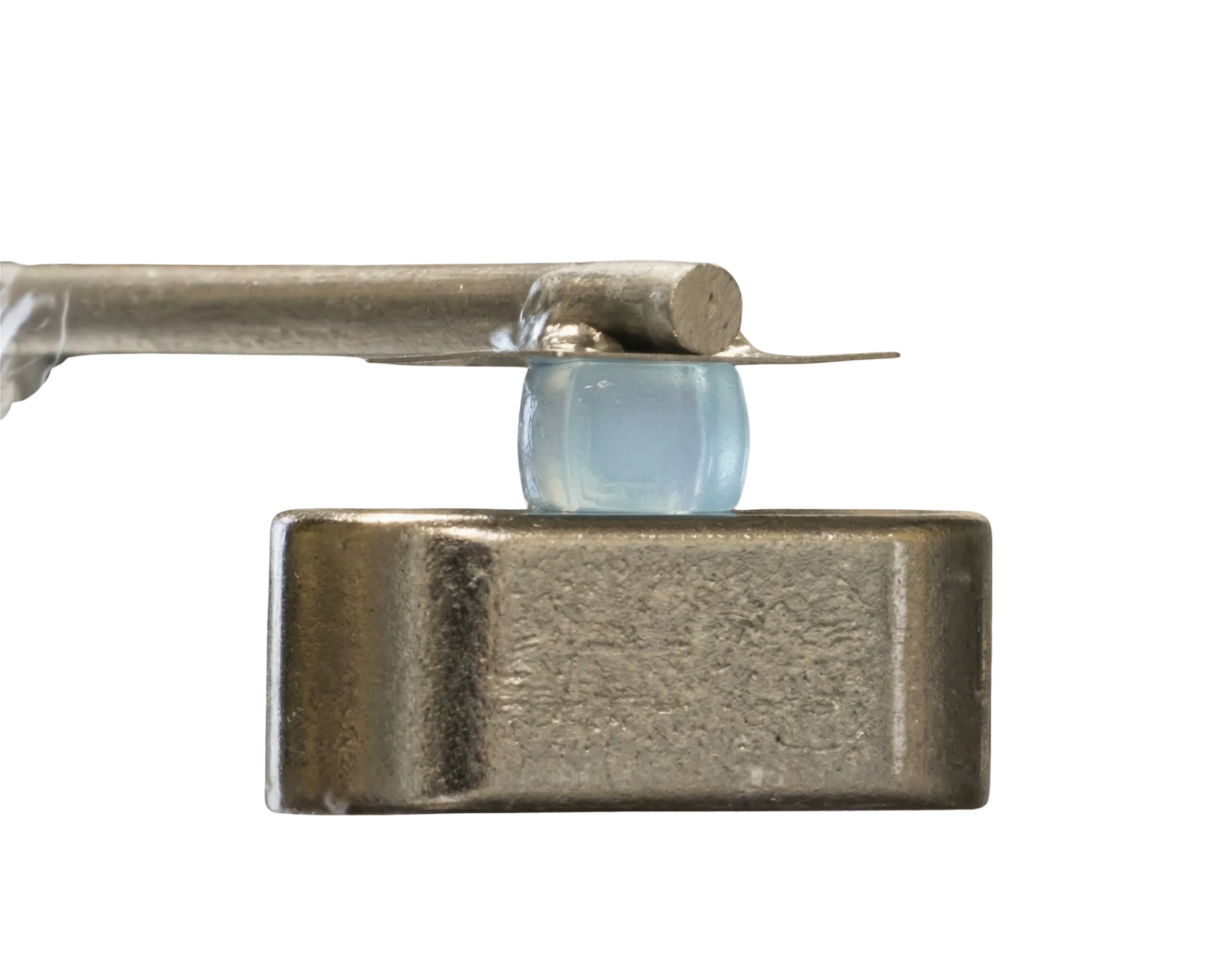
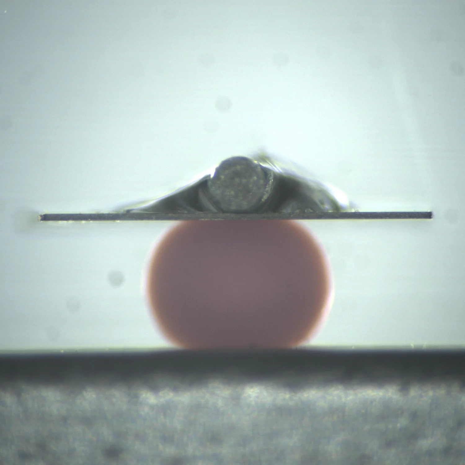
Mechanical Testing of Soft Gels Using the MicroTester
Demonstration of compression and indentation testing methods for soft gels using the MicroTester, showing consistent modulus measurements across geometries.
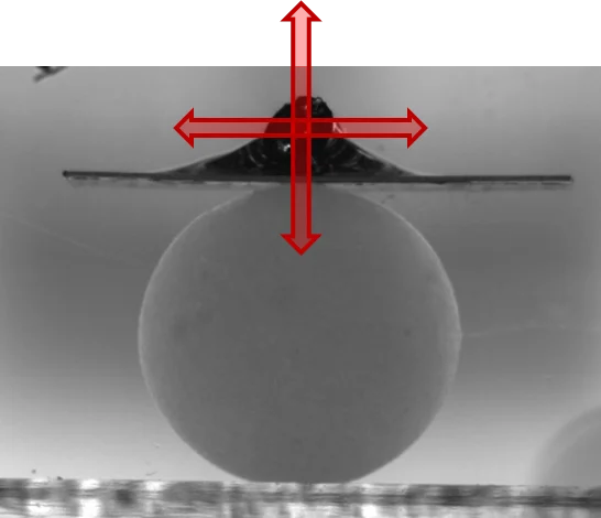
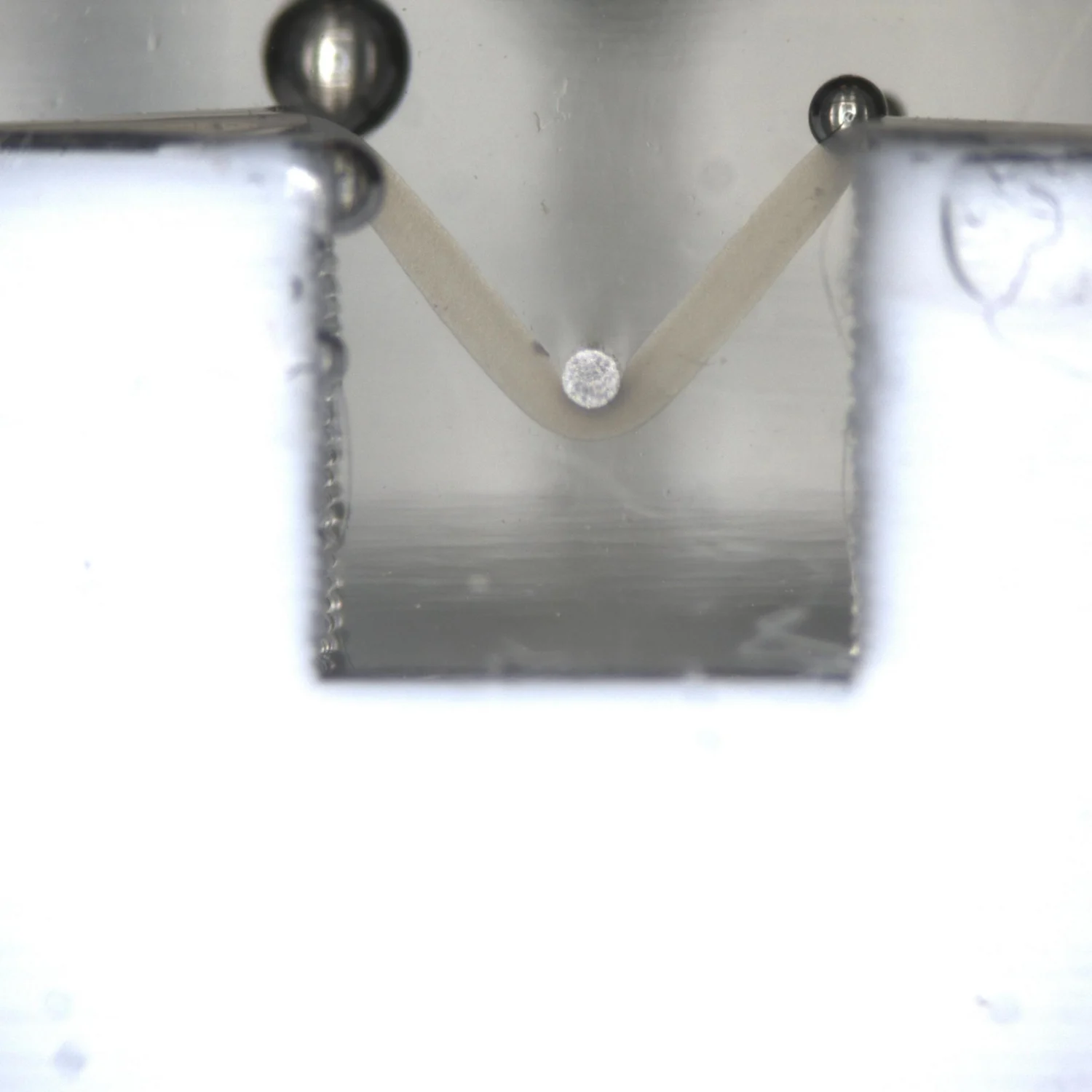
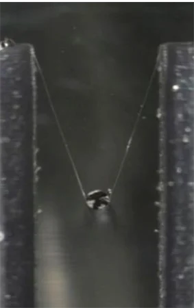
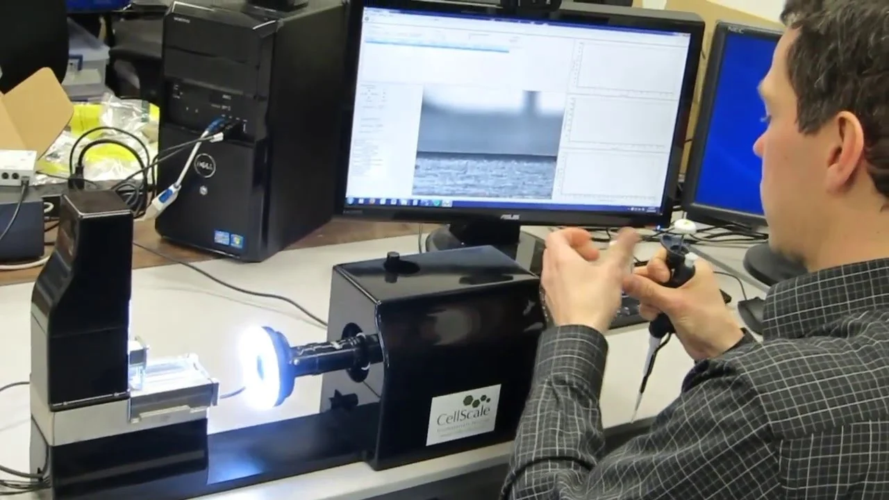
Micro-Scale Mechanical Testing Demonstration Using the MicroTester
Demonstration of micro-scale mechanical testing, highlighting compression, tension, and bending tests on ultra-small biomaterial specimens.
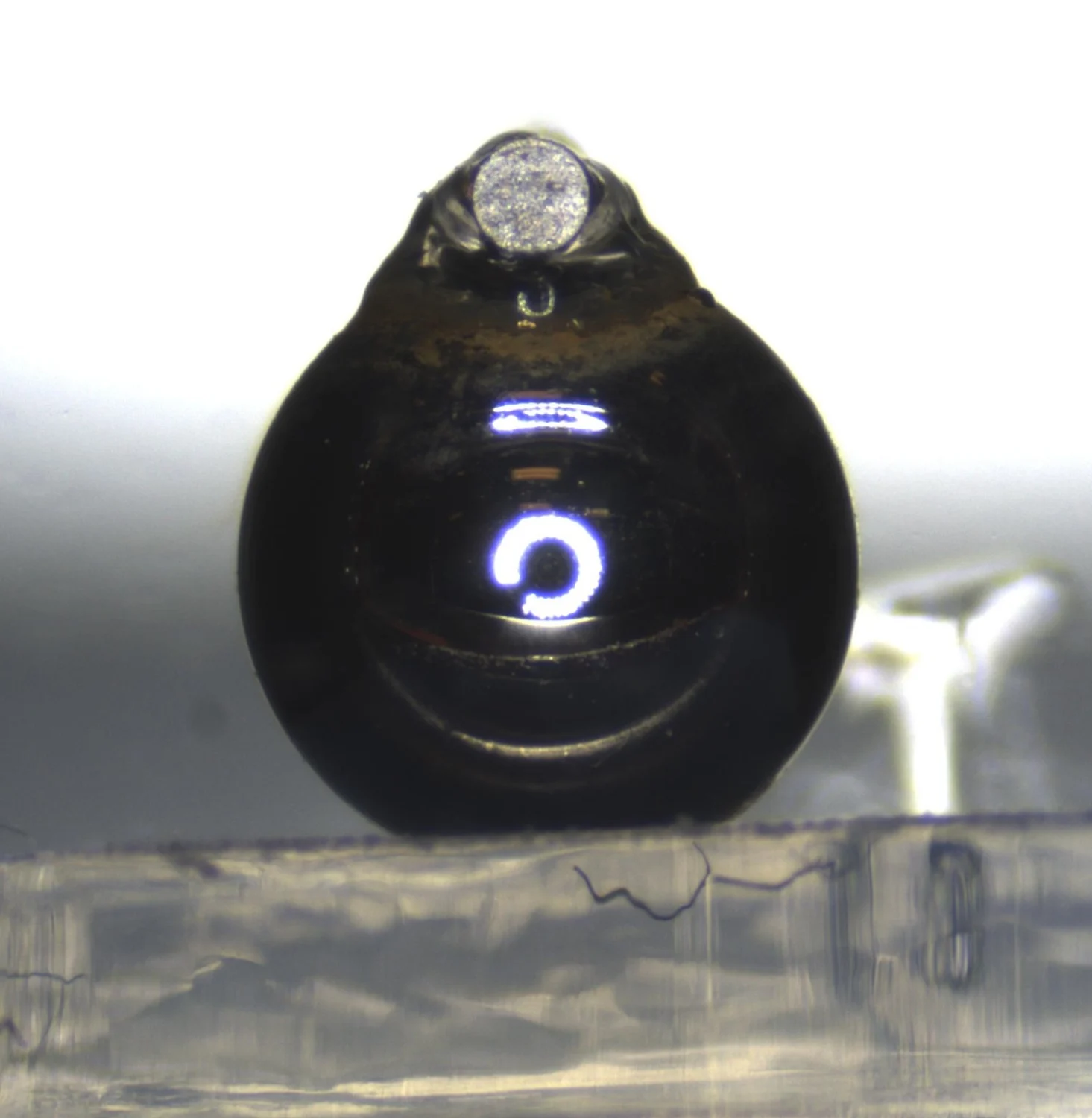
Mechanical Characterization of 3D Muscle Tissue Constructs | Kent State University
Compression testing of engineered 3D muscle tissue constructs to benchmark stiffness against native skeletal muscle.