by Steve Dragos | May 28, 2026 | Research Highlights
Eye drops have a short window to work. Once a formulation reaches the eye surface, blinking, tear turnover, and drainage begin removing it almost immediately. For drugs with poor solubility or limited penetration into ocular tissues, that short residence time becomes...
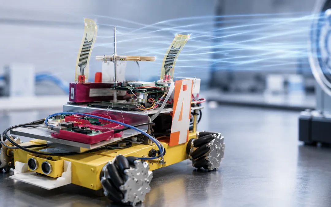
by Steve Dragos | May 14, 2026 | Research Highlights
Flexible sensors often look simple from the outside. A thin strip bends, a conductive layer changes resistance, and the signal is read by a circuit. In practice, especially in wearable bioelectronics and autonomous systems, that signal depends on a chain of mechanical...
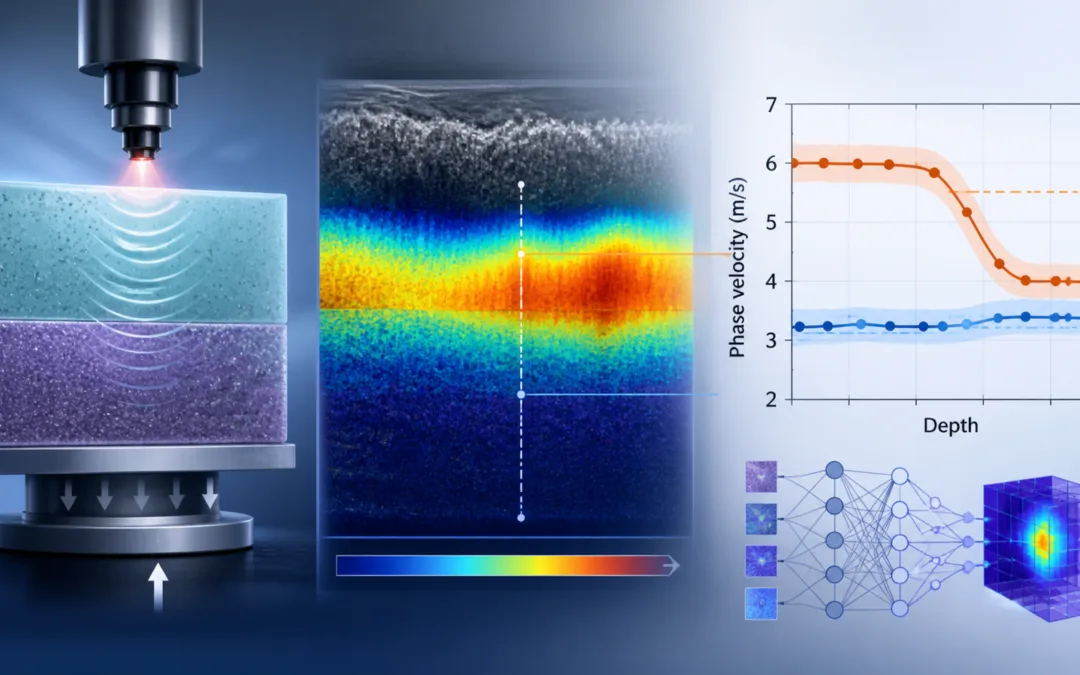
by Steve Dragos | May 7, 2026 | Research Highlights
Researchers are getting better at estimating tissue mechanics without cutting, stretching, or directly compressing the tissue being studied. Optical coherence elastography is one example. It uses optical coherence tomography to track wave motion through tissue, then...
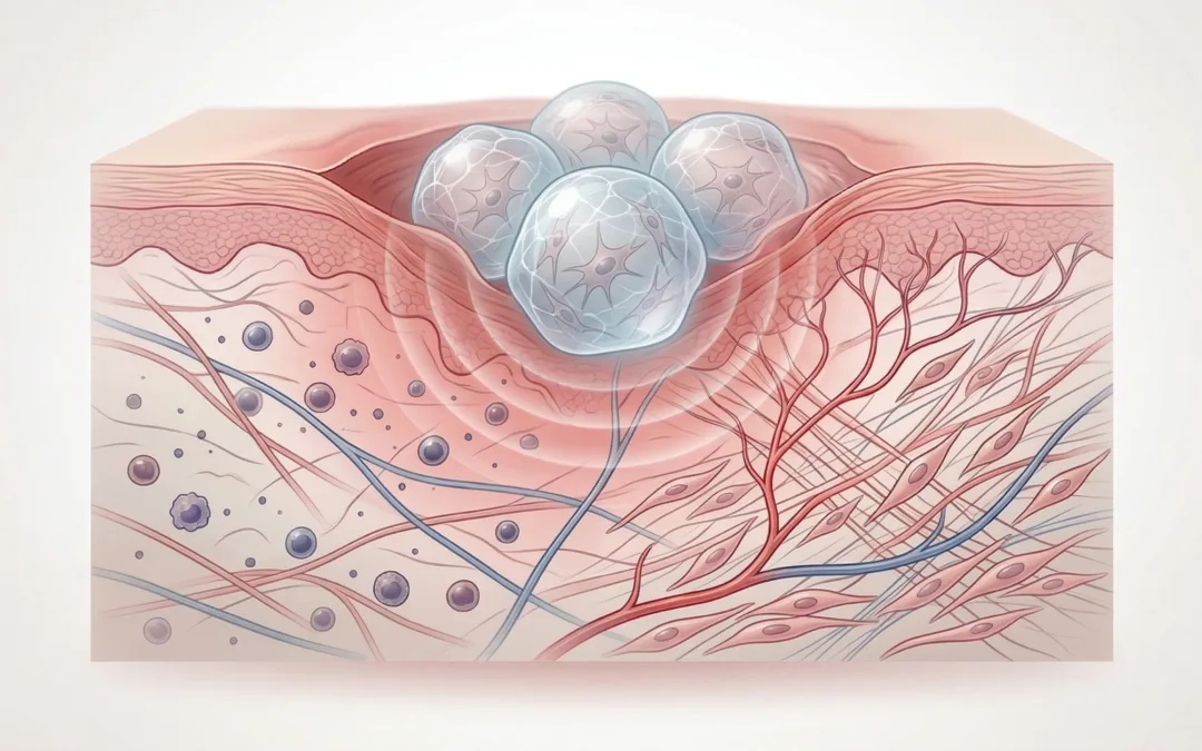
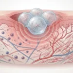
by Steve Dragos | Apr 30, 2026 | Research Highlights
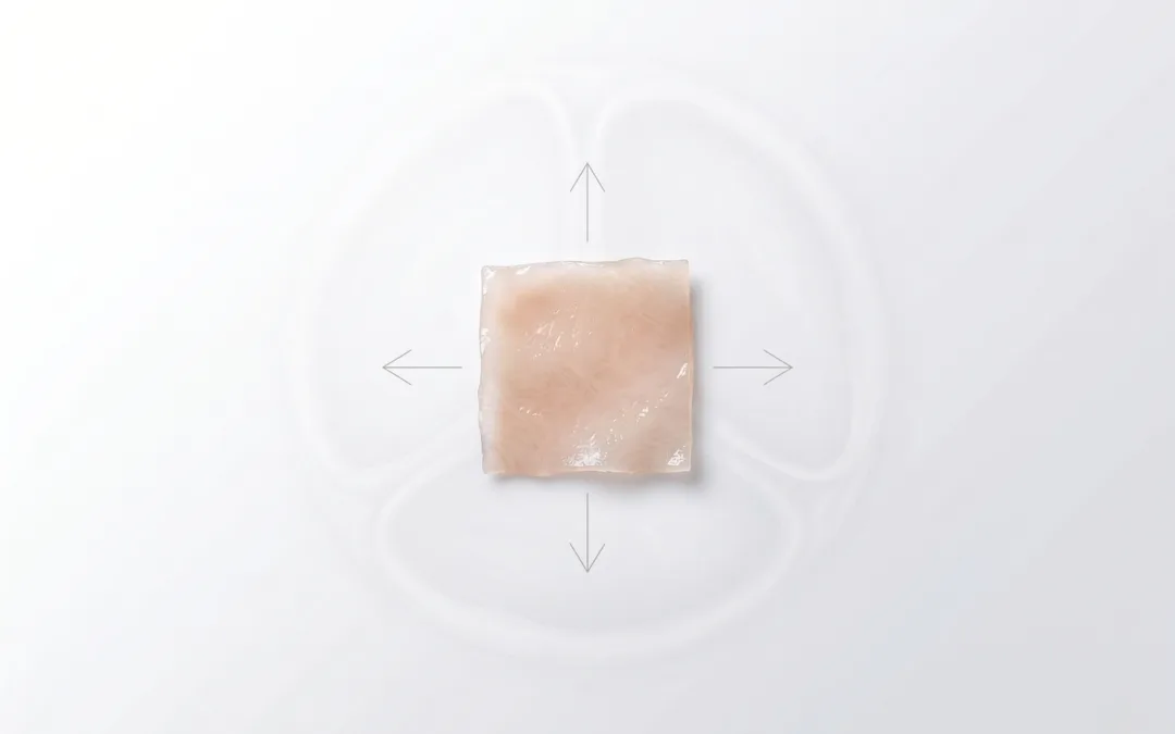
Chronic diabetic wounds are slow, but the deeper issue is that they stall. The inflammatory phase persists, angiogenesis stays weak, and remodeling does not kick in the way it should. That is part of what makes this study out of Sun Yat-sen University in China...
by Steve Dragos | Apr 23, 2026 | Research Highlights
Designing a durable bioprosthetic valve leaflet is not really about determining one impressive tensile number and moving on. In practice, the design process is messier than that. A leaflet opens and closes millions of times, sits in a controlled environment, and...