A study recently presented at the Orthopaedic Research Society (ORS) conference discussed finite element (FE) modeling of the rotator cuff. Understanding and treating rotator cuff (RC) tears, a common shoulder injury, can be challenging due to the complex structure of the supraspinatus tendon. This study aimed to assess the internal strains on small- and medium-sized RC tears under different conditions using the CellScale Univert system and FE modeling. Both tools are used to simulate the mechanical behavior of the tendon.

The study revealed that while tendon strain escalates with tear size, many daily activities and specific rehab exercises remain below the strain threshold that exacerbates tear progression, affirming their safety for those with RC injuries. The research suggests that the use of patient-specific FE modeling in clinical settings will help to better understand and manage the risk of RC tear progression, highlighting the importance of tailored rehabilitation exercises based on the size of the tear and the specific strains applied to shoulder tendons.
Understanding and Managing Pathology
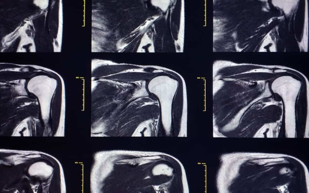
Shoulder pain stands out as a common issue, ranking third among musculoskeletal complaints in primary care settings. Rotator cuff (RC) tears, affecting around 23% of the population, are significant within this spectrum. While many RC tears may start symptom-free, eventually, 36% can become painful. Understanding and treating these tears effectively presents challenges due to the intricate structure and mechanics of the supraspinatus tendon. Finite element (FE) modeling offers researchers key insights into the mechanical behavior of this tendon, aiding in evaluating conditions that are hard to observe in vivo. The accuracy of these models in predicting tissue deformation is crucial for their clinical relevance. Tendons display a hyper-elastic response due to collagen fiber recruitment and alignment. This study’s core aim is to analyze internal strains in small- and medium-sized RC tears during typical activities and exercises to enhance the assessment of tear progression.
Characterization and Modeling of Supraspinatus Tendon Injuries
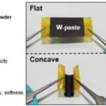
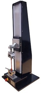
The procurement of six fresh-frozen intact human shoulders from Medcure, Inc. in Providence, RI, with an average age of 65 ± 9, was conducted with meticulous care. These shoulders underwent thorough examination to ensure the absence of rotator cuff tears, fraying, or delamination. Following the removal of skin and muscles, excluding the supraspinatus, infraspinatus, and subscapularis muscles, detailed isolation of the supraspinatus tendons was performed. Subsequently, the tendons were detached from the humeral head at the insertion site and meticulously divided into anterior, middle, and posterior regions, further segmented into medial and lateral regions as well as articular and bursal regions, yielding 12 samples per tendon, each measuring about 5 by 10 by 1 millimeters. Mechanical properties of each tendon region were assessed through uniaxial tensile testing using the UniVert system from CellScale BiomaterialsTesting in ON, Canada, up to 10% strain to prevent any permanent damage. Post mechanical testing, thin slices of 10 µm were obtained at intervals of 250 µm, stained with Masson’s trichrome, and imaged using a brightfield microscope (Olympus VS120) for collagen orientation evaluation.
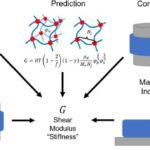
For the characterization of tendon regions for an FE model, collagen orientation and stress-strain curves served as inputs to fit the Holzapfel-Gasser-Ogden model for soft tissue, determining essential material parameters for FE modeling. By utilizing the established supraspinatus-infraspinatus model geometry, simulations of crescent-shaped tears were conducted in the posterior third of the supraspinatus tendon, a region linked to the origin of rotator cuff tears, encompassing small- and medium-sized tears.
The study aimed to precisely determine the percentage of maximum voluntary contraction (MVC) utilized during daily activities and rehabilitation exercises, focusing on the supraspinatus and infraspinatus muscles. This analysis provides comprehensive insights into muscle utilization patterns, enhancing understanding of movements.
Tendon Tear Analysis
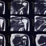
The maximum principal strains on the articular and bursal sides of the tendon were meticulously measured at the precise anterior and posterior edges of the tear tips as shown in Figure 1.
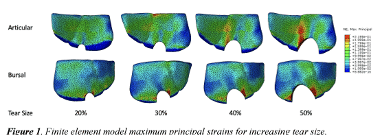
As the tear size increased, there was a corresponding rise in the strain experienced, but it consistently remained below the failure strain threshold indicating tear progression. An intriguing disparity emerged for smaller tears at 20% and 30%, where the bursal side showed higher strain levels at the anterior tip compared to the articular side. With progressing tear size, the articular side demonstrated more strain at the anterior tear tip during both prone abduction and external rotation. Insights from previous studies on cadavers suggested a strain percentage of 26.1 ± 9.4 as a failure threshold, highlighting a risk of tear progression for medium-sized tears during prone abduction at 40% and 50%. However, strain levels during external rotation remained below 26.1%, indicating a lower risk. Daily activities like typing, drinking, and brushing teeth kept strain levels below 16.7%, suggesting they are unlikely to induce tear progression.
Topic for Discussion:
The Finite Element (FE) modeling method provides vital insights into the intricate internal strains experienced by the rotator cuff (RC), revealing details that may elude in vivo measurements. Understanding these internal mechanics is crucial for early identification of patients prone to tear progression. Physical therapy, which boasts a remarkable 75% success rate in managing small- to medium-sized RC tears, faces challenges as tears worsen and symptoms persist. Tailoring rehabilitation protocols to the specific tendon strain and developing personalized care plans considering tear size and applied loads empower healthcare professionals to effectively mitigate tear progression risks and enhance patient outcomes. By customizing interventions to individual needs, healthcare providers can optimize the management of RC tears and improve patient recovery.
Patient-Specific Modeling and Rehabilitation Strategies
The application of patient-specific finite element modeling, involving the creation of virtual models tailored to each patient’s unique characteristics, proves to be clinically valuable in assessing tear progression risk. By considering individual material properties and specific tear features, this approach enhances the accuracy of predicting tear development over time. These research findings underscore the significance of integrating personalized assessments into current risk management strategies for minor Rotator Cuff tears.
However, to delve deeper into these insights, further exploration of tear characteristics and a comprehensive analysis of rehabilitation load applications for medium-sized tears are imperative. This enhanced comprehension will aid in identifying the most effective and tailored exercises to optimize patient outcomes and rehabilitation success.
Read more about it here.