Optic nerve head biomechanics play a central role in glaucoma research because elevated intraocular pressure places the optic nerve head under abnormal mechanical strain. Those deformations are thought to contribute to retinal ganglion cell damage, altered blood and nutrient supply, tissue remodelling, and progressive vision loss. For that reason, understanding the optic nerve head mechanical properties of relevant animal models is important for both experimental and computational glaucoma research.
This study from Elizabeth Boazak, Johan d’Humières, A. Thomas Read, and C. Ross Ethier focused on the compressive mechanical properties of optic nerve head tissue in rats and pigs. That focus is important because most published ONH mechanical data had previously come from tensile testing, even though compression is the main mode of deformation in the optic nerve head under elevated intraocular pressure. CellScale contributed to the work through the use of a CellScale MicroSquisher (now MicroTester), making this a useful example of how CellScale compression testing supports ocular biomechanics research.
Why compression testing matters for glaucoma biomechanics
One of the clearest strengths of this paper is its mechanical framing. The authors explicitly note that although tensile data are more common in the literature, compression testing of optic nerve head tissue is more physiologically relevant because compression is the predominant deformation mode in the ONH under elevated IOP. That makes this study much more than a material-property note. It is a methodological correction to how ONH biomechanics should be approached.
This is exactly why the paper matters for glaucoma biomechanics. If in vitro models and finite element simulations are built using the wrong loading assumptions, their mechanical predictions may be less relevant to the real tissue environment. By measuring ONH response in unconfined compression, the study gives researchers more appropriate inputs for model development.
For another ocular biomechanics application, see our post on corneal stiffening.
What the researchers tested
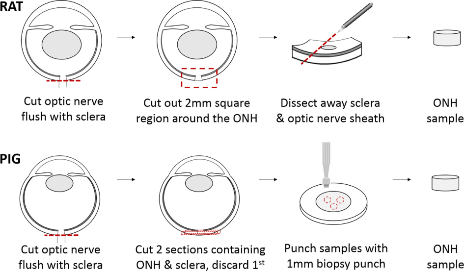
The study examined both pig and rat optic nerve head tissues, two species commonly used in glaucoma research. Pig ONH samples were punched from thin transverse sections containing the lamina cribrosa region, while rat ONH samples were carefully dissected to isolate the glial lamina. These species were a useful comparison because the pig ONH includes a collagenous lamina cribrosa, while the rat ONH has a non-collagenous glial lamina.
That species difference matters mechanically. It gave the authors a way to compare two distinct ONH tissue architectures under the same compression protocol, which helps explain why the resulting stiffness values were substantially different between pig and rat tissues.
How the CellScale MicroTester was used
The samples were tested in unconfined compression in PBS between 35 and 37C on the MicroTester. Compression was applied along the nerve axis, with pig samples preloaded to 50 mN and rat samples to 3 mN before cyclic loading. After preload, both tissue types were subjected to five cycles of unconfined compression to 15% compressive deformation at either 5% per minute or 40% per minute.
The neo-Hookean model provided species-specific material constants
To quantify the optic nerve head mechanical properties, the authors fit the loading portion of the stress-strain data to an incompressible neo-Hookean material model. The equilibrium material constant C1 was much higher in pig ONH than in rat ONH. At the 5% per minute strain rate, the mean C1 values were 428 Pa for pig ONH and 64 Pa for rat ONH.
That difference is one of the paper’s most important findings. The pig optic nerve head was substantially stiffer than the rat optic nerve head, which the authors interpreted as consistent with the pig’s collagenous lamina cribrosa compared with the rat’s glial lamina. For researchers building computational modeling of glaucoma workflows, that kind of animal-specific difference is exactly the sort of information that should not be ignored.
Rat tissues showed stronger strain-rate dependence
The paper also looked at how strain rate affected ONH behaviour. A subset of samples was tested at both 5% per minute and 40% per minute. In rat ONH, the C1 values increased significantly with the higher strain rate, from 81 Pa to 121 Pa. In pig ONH, the same trend was observed, but it was not statistically significant in this dataset.
This is a valuable point for optic nerve head biomechanics because the ONH is exposed not only to steady IOP but also to oscillatory loading associated with the ocular pulse. The authors note that physiological strain rates can be much higher than the ones used here, especially in rats, which means rate dependence could become even more relevant in future model systems.

Why these data matter for glaucoma models
The broader value of the study is that it gives researchers better mechanical inputs for species-specific glaucoma models. The authors explicitly state that the reported compressive properties should help improve finite element models of the ONH and support in vitro platforms for studying astrocyte and retinal ganglion cell mechanobiology. Because neo-Hookean material models are readily available in most finite element packages, these values are easy for other groups to use.
That makes this paper especially useful for engineering audiences. It is not just about describing tissue stiffness. It is about creating better computational modeling of glaucoma and more realistic in vitro environments for ONH cell biology.
Why this matters for animal-specific ocular biomechanics
A key takeaway is that rat and pig optic nerve heads should not be treated as mechanically interchangeable. The paper shows clear species-level differences in compressive stiffness, which likely reflect structural differences between the collagenous porcine lamina cribrosa and the glial rat lamina. That is important when choosing animal models and when interpreting how well a given model reflects the human eye.
Final thoughts
This study is a strong example of how optic nerve head biomechanics can be approached in a more physiologically relevant way. By focusing on compression rather than tension, the authors measured the mechanical response of rat and pig ONH tissues under a loading mode that better reflects elevated intraocular pressure. They also showed clear species differences, meaningful strain-rate dependence in rat tissue, and a straightforward path toward model integration using neo-Hookean parameters.
For CellScale, the paper highlights the value of compression-based testing for soft ocular tissues. The MicroTester supported the measurements that linked tissue architecture to material response, making this a useful research highlight for both glaucoma audiences and users interested in microscale soft tissue mechanics.
Read the full journal article here: Compressive mechanical properties of rat and pig optic nerve head
Read about Dr Ethier’s research here: Ethier Lab
For related reading, you may also like: