Age-dependent skin mechanics matter in far more than basic biomechanics. They influence how skin responds to loading, how it changes with aging, how wounds close, how tissue is modelled in simulation, and how researchers interpret cosmetic or reconstructive studies. Because mouse models are widely used in skin biology, having accurate mechanical data for murine skin is important for both experimental interpretation and predictive modelling.
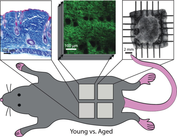
In this study, researchers at the University of Texas at Austin carried out a detailed comparison of young and aged mouse skin using histology, 2-photon microscopy, and planar biaxial testing. CellScale contributed to the work through the use of the BioTester, which was used to quantify the biaxial response of skin under controlled loading. The result is one of the most complete datasets on age-dependent skin mechanics in mouse skin, linking structure, collagen organization, residual strain, and constitutive behaviour in a way that is especially useful for computational biomechanics.
For broader context on soft tissue characterization, see our guide to mechanical testing of biomaterials.
Why age-dependent skin mechanics matter
Skin is a nonlinear, anisotropic soft tissue whose behaviour depends on collagen architecture, elastin contribution, tissue layering, hydration, and loading direction. Those properties also change with age. For that reason, age-dependent skin mechanics are relevant not only to skin biology, but also to wrinkle formation, wound healing, surgical planning, and computational modelling.
The authors make this point clearly. Mouse skin is widely used as a model for human skin in cosmetic science and medical science, but the literature still had major gaps around region, age, direction, and biaxial behaviour. This study was designed to fill those gaps by comparing young and aged murine skin across dorsal and ventral regions while also accounting for residual strain and constitutive modelling.
What the researchers measured
The study combined three main approaches. Histology was used to quantify layer thickness and tissue composition. 2-photon microscopy was used to assess collagen fiber orientation through the dermis. Planar biaxial testing was then used to measure the mechanical response of the tissue in lateral and cranial-caudal directions.
That combination is what makes the paper especially strong. Rather than reporting only mechanical curves, the authors connected biaxial testing of skin tissue to actual microstructural and regional differences in the skin. This gives the work much more value for readers interested in mouse skin biomechanics and computational modeling of skin.
How the CellScale BioTester was used
CellScale’s role in the study came through the BioTester, which the team used for planar biaxial mechanical testing. Samples from dorsal and ventral skin were prepared, speckled for optical tracking, preloaded, and then tested under off-biaxial and equibiaxial loading protocols in PBS at 37 °C. The recorded data were later used to calculate strain, stiffness, stretch, and constitutive model parameters.
This is an important detail because skin is not well represented by a single uniaxial number. Biaxial testing of skin tissue is much more informative for a material that naturally experiences multi-directional loading and anisotropic fibre recruitment. In this paper, BioTester-based testing helped reveal directional differences in stiffness and stretch response that would be difficult to capture with simpler testing methods.
For another example of biaxial soft tissue mechanics, see our post on venous valve tissue mechanical properties and ECM.
Histology showed structural differences with aging
The histology results showed that skin structure changed with age, particularly across the dermis and hypodermis. Dermal thickness decreased with age, while the hypodermis became thicker in aged mice, especially in ventral skin. The authors did not observe major changes in epidermal thickness or collagen area fraction, but the layer-level structural changes were still important because they help explain why age-related skin behaviour cannot be reduced to a single simple metric.
This matters for age-dependent skin mechanics because tissue composition and thickness directly influence when and how a sample begins to stiffen during loading. It also reinforces the translational angle of the paper. Aging skin is not only a cosmetic question. It is a structural and mechanical question.
2-photon microscopy linked collagen orientation to anisotropic skin behaviour
The 2-photon microscopy analysis showed that collagen fibers were broadly distributed but had a mean orientation that generally aligned laterally. That is important because the skin also showed greater stiffness in the lateral direction during biaxial testing. In other words, the structural observations and the mechanical results pointed in the same direction.

This is one of the strongest sections of the study because it supports anisotropic skin behaviour with both imaging and mechanics. The authors also found that collagen dispersion changed with depth, which adds another useful layer for readers interested in multiscale skin mechanics and constitutive modelling.
Residual strain changed the interpretation of skin behaviour
A major contribution of the paper is its analysis of residual strain. The authors showed that ventral skin was under notable residual strain in situ, while dorsal skin showed much less. That finding matters because neglecting residual strain can change how stress-stretch behaviour is interpreted and can shift constitutive curves in ways that matter for simulation.
This is especially important for computational modeling of skin. If the reference configuration is chosen incorrectly, the model may still look mathematically reasonable while being mechanically misleading. The study explicitly shows that the constitutive response changes depending on whether the tissue is referenced to the preloaded in vitro, unloaded in vitro, or unloaded in situ configuration.

For readers interested in another viscoelastic or reference-state problem in soft tissues, see our post on biomechanical analysis of the urinary bladder.
Biaxial testing showed directional and regional differences
The biaxial results confirmed that mouse skin is anisotropic. Toe-region stiffness was higher in the lateral direction than in the cranial-caudal direction. The tissue also tended to stiffen earlier on the dorsal side than the ventral side. These findings support the idea that mouse skin behaviour depends not only on age, but also on region and direction.

The aging effect itself was more subtle than some readers might expect. The study found that aged skin became more compliant in terms of the stress-stretch curve shifting toward larger stretches, particularly on the ventral side, but the tangent modulus at higher stretch did not change dramatically. That nuance is useful because it avoids oversimplifying skin aging into a single statement about “stiffer” or “softer” skin.
Why this dataset matters for computational modeling of skin
One of the strongest messages in the paper is that the dataset is not just descriptive. It is built to support modelling. The authors used the measured biaxial response to inform a Fung-type hyperelastic constitutive law, allowing the structural and mechanical data to be translated into parameters that can be used in predictive simulations.

That makes the study particularly useful for engineering audiences. It provides not only histology and microscopy observations, but also constitutive descriptions that can be used in finite element or other computational workflows. For anyone working on computational modeling of skin, this is one of the most practical reasons to read the paper.
Final thoughts
This study is one of the strongest examples of age-dependent skin mechanics in murine tissue because it combines histology, collagen imaging, residual strain analysis, and BioTester biaxial testing in a single framework. It shows that mouse skin is anisotropic, region-dependent, and sensitive to reference configuration, while age introduces structural and mechanical changes that are meaningful but not overly simplistic.
For CellScale, the study also highlights the value of the BioTester in biaxial testing of skin tissue. The instrument supported the mechanical measurements that tied skin structure back to directional behaviour and constitutive modelling, which is exactly the kind of workflow researchers need when moving from tissue characterization to predictive simulation.
Read the full journal article here: The regional-dependent biaxial behavior of young and aged mouse skin: A detailed histomechanical characterization, residual strain analysis, and constitutive model
Read about Dr. Rausch’s research here: Manuel Rausch Lab
For related reading, you may also like: