Understanding urinary bladder mechanical properties is important for the development of new therapies for stress urinary incontinence, pelvic floor repair, implantable stimulators, and engineered tissue replacements. The bladder is not just a passive storage organ. Its wall shows time-dependent soft tissue behaviour, and that mechanical response matters when researchers want to model function, design devices, or build realistic tissue substitutes.
In this study, researchers from Tecnologico de Monterrey and the University of Texas at Dallas combined 3D surface reconstruction with tensile testing to evaluate bladder tissue mechanics. CellScale contributed to the work through the use of the UniVert, which was used to generate stress-strain data from bovine bladder samples. Those measurements were then used to estimate tissue elasticity and damping through a linear viscoelastic Voigt model. This gives the paper value for both biomechanical testing of bladder tissue and viscoelastic modeling of soft tissues.
For broader context on why these measurements matter, see our guide to mechanical testing of biomaterials.
Why urinary bladder mechanical properties matter
The bladder must accommodate volume changes while maintaining functional control over pressure and deformation. That makes it a mechanically interesting tissue, especially in the context of disorders such as stress urinary incontinence. For device developers and tissue engineers, knowing the urinary bladder mechanical properties of the organ wall is a necessary first step toward more realistic models and better treatment strategies.
This is where the present study is useful. Instead of looking only at tissue samples or only at geometry, the authors combined physical surface capture with tensile characterization. That pairing makes the work relevant to researchers interested in both organ-level reconstruction and material-level response.
What the researchers set out to measure
The team focused on two related goals. First, they created a digital surface representation of the bovine urinary bladder using 3D scanning. Second, they performed tensile testing on tissue taken from different bladder regions so they could estimate elastic and viscous parameters with a Voigt model.
That combination is important because it links shape and mechanics. The resulting workflow supports not only biomechanical testing of bladder tissue, but also future computational modelling where realistic organ geometry and material behaviour need to work together.
How the urinary bladder surface was reconstructed
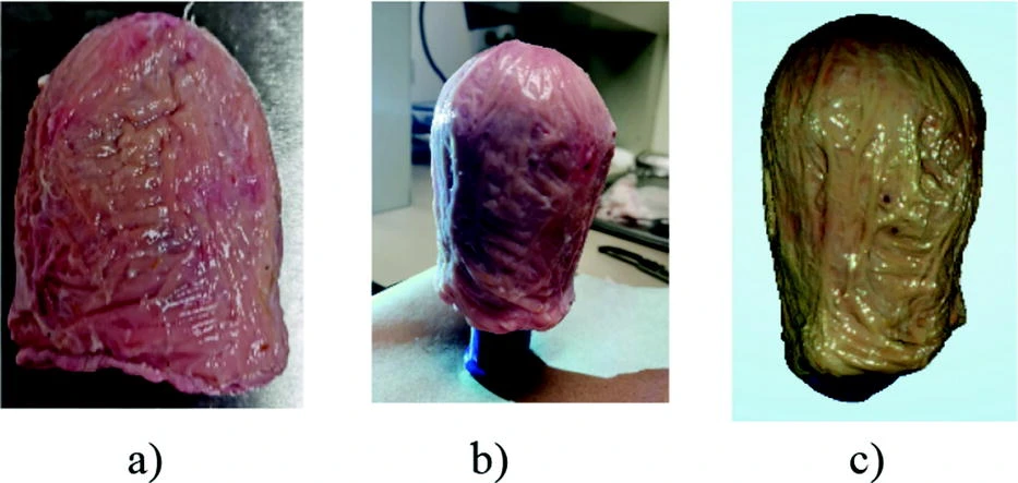
The researchers obtained bovine bladders and prepared them for 3D scanning after cleaning and hydration. The organ was scanned and reconstructed as a point cloud and triangular mesh, creating a digital bladder surface with a very high point count and mesh density. The reconstructed model was then used as the basis for a more complete surface representation of the organ.
This part of the study is useful for readers interested in urinary bladder biomechanics, because it shows how tissue characterization can be paired with digital organ capture. The geometry work is not just visual. It supports later computational use.
How the CellScale UniVert was used
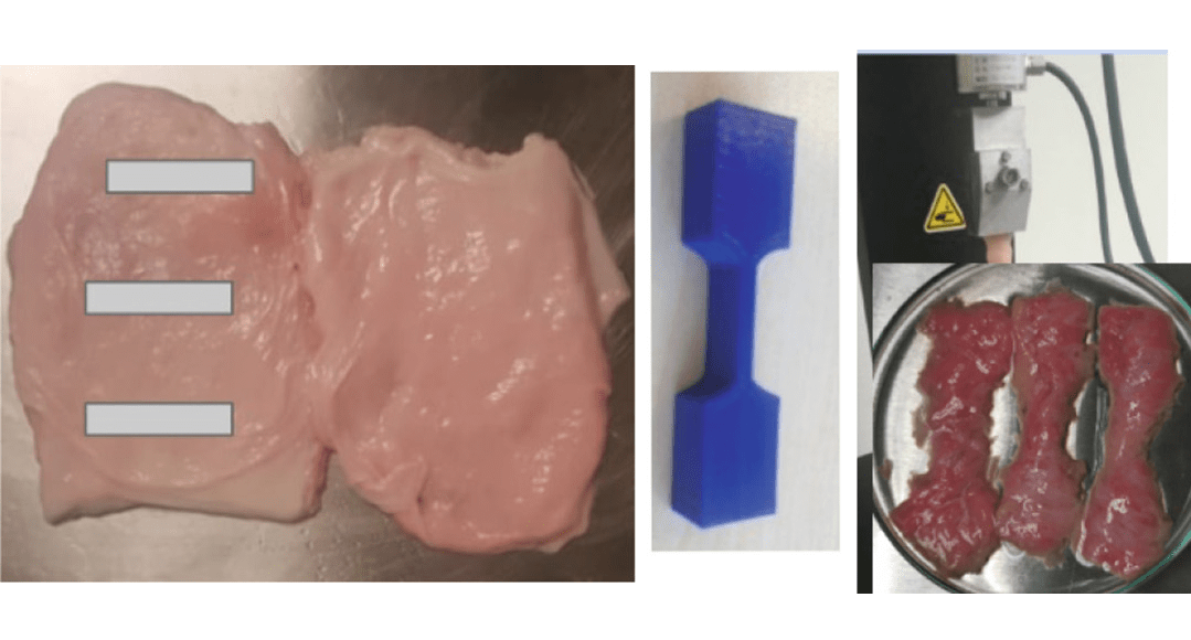
CellScale’s role in the study came through the UniVert, which was used to perform tensile testing on bladder tissue samples. After surface reconstruction, the researchers divided the bladder into upper, middle, and lower regions, then cut tissue specimens using a printed dogbone-style template to standardize sample dimensions.
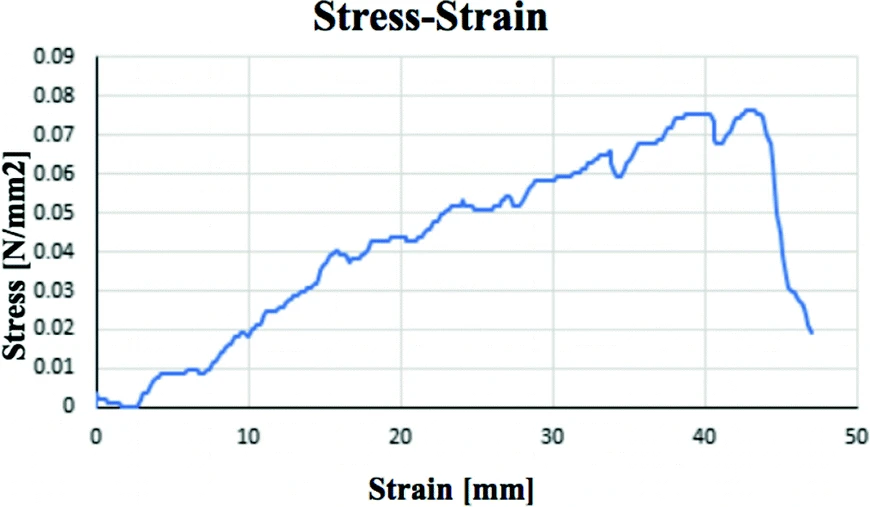
The UniVert system was used with a 200 N load cell to apply tensile loading and generate stress-strain data from the bladder wall samples. That mechanical data formed the basis for estimating material parameters in the viscoelastic model. This is an important part of the story because it places CellScale directly in the transition from tissue sampling to quantitative mechanical characterization.
For another example of regional tissue mechanics, see our post on regional variances in muscle tissue.
Why tensile testing of bladder tissue was important
The core experimental step in the paper was tensile testing of bladder tissue from different anatomical regions. By testing upper, middle, and lower bladder samples separately, the researchers could compare whether the tissue behaved uniformly or whether some regions were stiffer or more viscous than others.
That regional view matters because soft tissues are often mechanically heterogeneous. A single average value may not describe the whole organ well enough for simulation or device design. In this study, the regional testing approach showed that bladder mechanics varied across the tissue, which strengthens the case for more detailed modelling rather than assuming a single uniform material response.
Voigt model analysis and viscoelastic tissue behaviour
A key strength of the paper is that it goes beyond simple tensile results and explicitly frames the tissue as viscoelastic. The researchers used a linear dynamic mechanical model based on Voigt’s formulation to estimate two main parameters: elasticity and damping.
That is useful because viscoelastic modeling of soft tissues is exactly the kind of language that better matches how this study is positioned scientifically. The tissue was not treated as purely elastic. Instead, the stress-strain data were used to identify an elastic constant and a viscous constant, giving a more realistic description of bladder wall response.
For a related method-focused article, see viscoelasticity testing.
What the study found about bladder tissue mechanics
The authors found regional differences in bladder wall behaviour. The lower region had the greatest elastic response, while the upper region showed the strongest viscous response. Average values for elasticity and viscosity were also reported across the sampled tissues, supporting the idea that the bladder cannot be fully described as a mechanically uniform structure.
This is one of the most useful takeaways from the study. For researchers working on urinary bladder mechanical properties, it suggests that both anatomy and region matter when interpreting tissue data. For engineers building computational or device-based models, that is an important reminder that simplification has limits.
Why this matters for stress urinary incontinence and med-tech design
The clinical framing here is worth emphasizing. Better knowledge of urinary bladder mechanical properties can support the design of therapies for stress urinary incontinence and related pelvic floor problems. It can also help guide work on artificial tissues, surgical repair strategies, and implantable devices that interact with bladder tissue.
The paper is therefore useful not only as a characterization study, but also as a supporting asset for med-tech and implant design audiences. It provides a practical example of how organ geometry, tissue testing, and viscoelastic modelling can be brought together in a single workflow.
Final thoughts
This study is a strong example of how urinary bladder mechanical properties can be investigated through both digital surface reconstruction and experimental tissue mechanics. By combining 3D scanning, regional tissue sampling, UniVert tensile testing, and Voigt model analysis, the researchers created a workflow that is useful for both organ modelling and soft tissue characterization.
CellScale’s UniVert played a direct role in the study by generating the tensile data used to estimate elasticity and damping. That makes this paper especially relevant for readers interested in biomechanical testing of bladder tissue, viscoelastic modeling of soft tissues, and translational soft tissue mechanics.
Read the full journal article here: Surface Representation and Bio-mechanical Analysis of the Urinary Bladder
Read more about Dr. Garcia-Gonzalez’s research here: Tecnologico de Monterrey research profile
For related reading, you may also like: