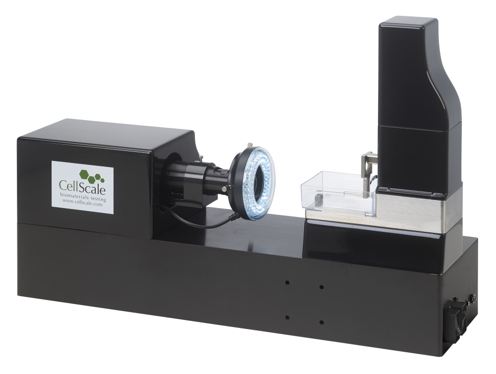
MicroTester
Superior Micro-Scale mechanical testing
Enhanced for smaller samples, the MicroTester offers improved force resolution, streamlined test setups, and exceptional visual feedback. It’s ideal for a wide range of applications, from tiny tissue samples to testing the properties of hydrogel microspheres, cell spheroids, and engineered microtissues.
MicroTester G2
- Compression, tension, bending, indentation and shear testing
- Piezo-electric actuators with 0.1µm resolution
- Optional second axis imaging
- Force resolution down to 10nN
- High resolution CCD imaging
- Integrated temperature-controlled media bath
- Fully featured user interface software for simple, cyclic, relaxation, and multi-modal testing with real-time feedback
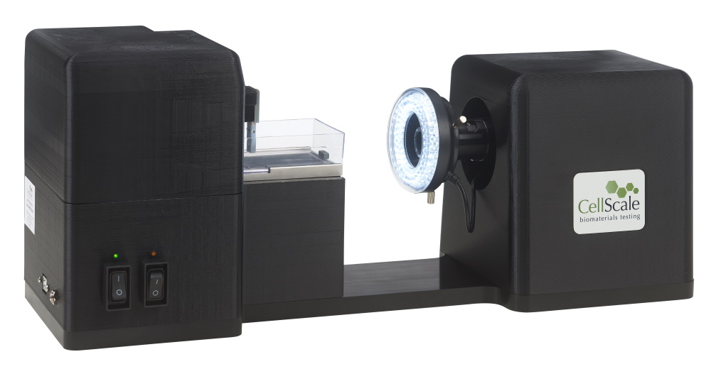
MicroTester LT
- Compression, tension, bending and indentation testing
- Affordable pricing for a wide range of applications and users
- Stepper motor actuators with 1µm resolution
- Force resolution down to 50nN
- High resolution CCD imaging
- Integrated temperature-controlled media bath
- Fully featured user interface software for simple, cyclic, relaxation, and multi-modal testing with real-time feedback
TECHNICAL INFO

| MTG2 | MTLT | |
| Dimensions | 56 X 14 X 24cm | 52 X 17 X 21cm |
| Weight | 9kg | 6.5kg |
| Force Capacity | 500mN | |
| Available Force Transducers | 0.005, 0.02, 0.08, 0.2, 1, 5, 25, 100, 500mN | |
| Force Accuracy | Approx. 0.2% of transducer capacity | |
| Maximum Grip Separation | Approx. 10mm | |
| Maximum Velocity | 0.5mm/s | |
| Maximum Cycle Frequency | 0.5Hz | 0.1Hz |
| Maximum Data Rate | 15Hz | 5Hz |
| Actuator Technology | Piezo-electric Motor | Stepper Motor |
| Actuator Resolution | 0.1um | 1um |
| Range of Field of View | 0.4-11.0mm | 0.8-5.5mm |
| Vertical Image Resolution | 2048px | 1536px |
| Secondary Camera Option | Yes | No |
| Secondary Test Axis Option (Shear) | Yes | No |

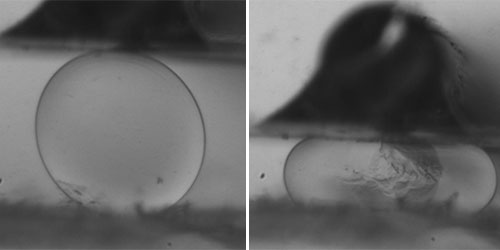
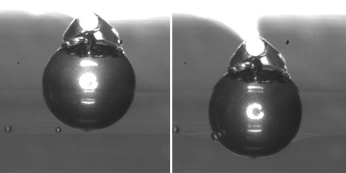
Specimens & Mounting
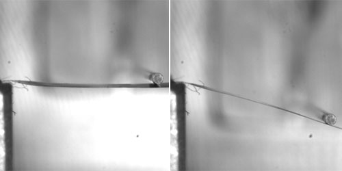
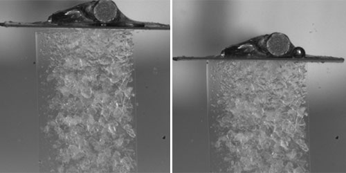
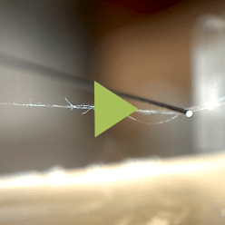
Bend-Induced Tension Specimen: spider silk Peak Force: 2mN
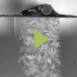
Puncture-Attached Tension Specimen: 3.5mm wide by 1.5mm thick hydrogel (0.5KPa) Peak Force: 1.4mN
Will The MicroTester Work For You?
The CellScale Microtester offers precision and versatility for applications ranging from biomaterial evaluation to micro-scale material testing. Its advanced features, including high-resolution imaging and dynamic mechanical analysis, provide essential insights into the mechanical behavior of a wide array of materials. Whether for academic research or industrial innovation, the Microtester delivers accurate, relevant data across diverse testing environments. Explore our whitepapers or contact our team today to see how the Microtester can meet your specific needs.
Get more information on the MircoTester
Enter your email below and we'll send you 3 case studies.
Videos
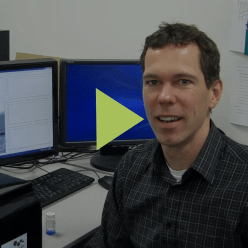
Our lab focuses on micro and nanoscale 3D-printing of soft hydrogels for tissue engineering applications. The MicroTester is a unique device capable of accurately testing our 3D-printed scaffolds where other methods have fallen short. The software provided is easy to use and provides flexibility for the diverse needs of our lab
CellScale MicroTester represents a unique opportunity to perform mechanical assays in cell spheroids together with a versatile optical system. We also have a great experience with CellScale’s customer support. See publication: Delivery of Human Adipose Stem Cells Spheroids into Lockyballs.
CellScale’s innovative equipment has allowed us to accurately characterize the properties of a diverse range of engineered bioscaffolds under physiological conditions. What truly sets CellScale apart, is their commitment to Customer Service – they have gone above and beyond to help us in getting the most from our data and are always very prompt and helpful in responding to our questions.
Publications

DOWNLOADS
If you would like help with updating your device software, please contact the CellScale team for free support.